
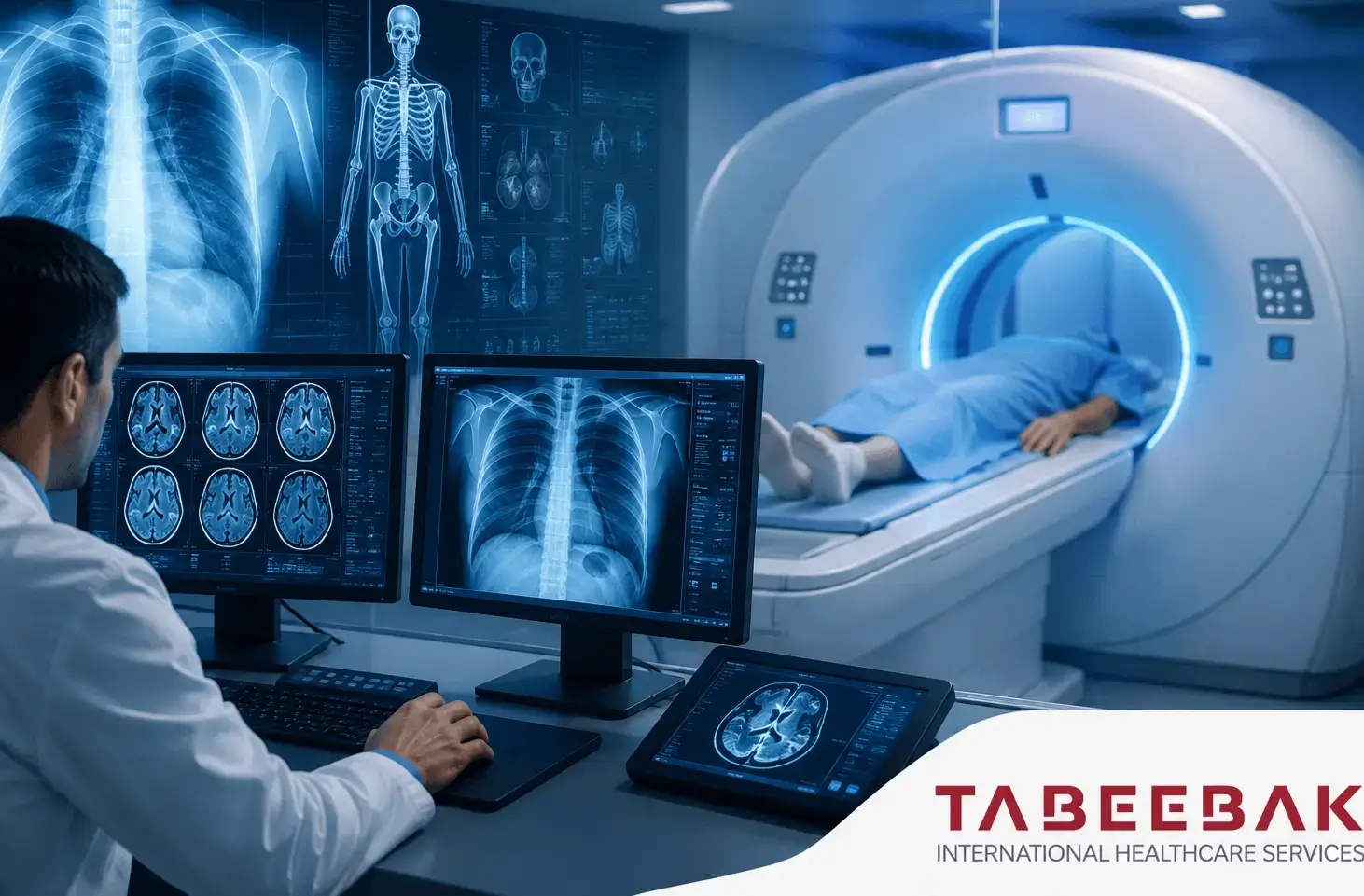
A doctor may request MRI, CT, or Ultrasound imaging depending on the organ and medical issue that needs to be evaluated. The newest or most expensive test is not necessarily the most appropriate one, as each imaging technique has different capabilities for visualizing tissues, bones, blood vessels, and internal organs.
The treating physician and radiologist in the Radiology and Medical Imaging Department determine the appropriate examination and imaging protocol based on the clinical question, the patient’s age, previous imaging studies, and overall health. The ACR Appropriateness Criteria provide evidence-based guidance for selecting the most appropriate imaging studies for different clinical situations.
What Is Radiology?
Radiology and Medical Imaging is the medical specialty that uses imaging technologies to visualize organs, tissues, bones, and blood vessels in order to support diagnosis, determine the extent of certain diseases, and monitor changes during treatment.
The role of a radiologist is not limited to interpreting images after they have been obtained. The radiologist may also help determine the appropriate imaging study and protocol, whether contrast material is needed, compare the examination with previous imaging, and provide conclusions that help the treating physician decide on the next step.
For this reason, radiology reports may differ even when the same body area is imaged if the clinical question is different.
What Is the Difference Between Diagnostic Radiology, Interventional Radiology, and Nuclear Medicine?
These specialties use imaging or radiation technologies, but they do not serve the same purpose.
| Specialty | Primary Role |
|---|---|
| Diagnostic Radiology | Imaging body organs and structures to support diagnosis and follow-up. |
| Interventional Radiology | Using imaging guidance to direct needles or catheters during diagnostic or therapeutic procedures. |
| Nuclear Medicine | Using radioactive substances for functional imaging and certain treatments. |
| Radiation Oncology | Using therapeutic doses of radiation to treat cancer and selected other conditions. |
For this reason, the term “radiology” should not be used to refer to all of these fields as if they were a single specialty. A patient who needs an MRI, for example, follows a completely different pathway from a patient who requires radiation therapy for cancer.
What Are the Main Imaging Tests in the Radiology and Medical Imaging Department?
The most appropriate imaging method depends on the organ and the condition being evaluated.
| Examination | Common Uses |
|---|---|
| X-ray | Bones, fractures, chest imaging, and certain initial evaluations |
| Ultrasound | Abdomen, liver, kidneys, thyroid gland, and superficial tissues |
| Doppler Ultrasound | Evaluating blood flow in arteries and veins |
| CT | Chest, abdomen, emergency imaging, bones, and blood vessels |
| MRI | Brain, spine, joints, and soft tissues |
| Mammography | Breast imaging and detection of certain abnormalities |
| Fluoroscopy | Real-time imaging of certain organs and procedures |
| CT Angiography | Imaging arteries and blood vessels |
These common uses do not mean that one imaging test is suitable for every problem involving the same organ. The choice depends on the specific clinical question the doctor needs to answer.
Magnetic Resonance Imaging (MRI)
MRI uses a strong magnetic field and radio waves to produce detailed images of organs and tissues. It does not use X-rays or ionizing radiation.
Magnetic Resonance Imaging is widely used to examine the brain, spinal cord, spine, joints, and muscles. Specialized MRI protocols are also available for imaging the heart, breast, abdomen, and pelvis.
MRI may be performed with or without contrast material. This depends on the reason for the examination and what the physician needs to assess, so not every MRI requires contrast injection.
When Might a Doctor Request an MRI?
MRI may be used to evaluate conditions involving:
- Brain and nervous system disorders.
- Certain tumors.
- Spinal disorders.
- Ligament and joint injuries.
- Certain heart conditions.
- Liver and pelvic disorders.
- Breast evaluation in selected cases.
Because MRI uses a strong magnetic field, the medical team must be informed about implanted devices or metal within the body before the examination so their MRI compatibility can be confirmed. The current MRI safety guidance emphasizes the importance of screening for risks related to implanted devices and metallic objects.
Does a Child Need Sedation or Anesthesia During MRI?
Not always. However, infants and some young children may require sedation or anesthesia if they cannot remain still throughout the examination. The decision depends on the child’s age, developmental level, and the type of imaging study.
Computed Tomography (CT)
Computed Tomography (CT) uses X-rays to create detailed cross-sectional images of the body. It can provide information about bones, organs, tissues, and blood vessels more quickly than some other imaging techniques.
CT is commonly used to image the chest, abdomen, and pelvis and to evaluate injuries, certain emergency conditions, tumors, and blood vessels.
A CT examination may be performed:
- Without contrast material.
- With intravenous contrast.
- Using a specialized protocol for a particular organ.
- As CT Angiography for blood vessel imaging.
CT or MRI?
There is no single answer that applies to every condition:
- CT is faster in certain emergency situations and may be particularly useful for the lungs, bones, bleeding, and some injuries.
- MRI provides different and often more detailed information about the brain, soft tissues, joints, and spinal cord.
Therefore, moving from CT to MRI should not be considered an “upgrade.” According to RadiologyInfo – Body CT, each examination answers different medical questions.
Ultrasound
Ultrasound uses sound waves to produce images of different parts of the body and does not use ionizing radiation.
It may be used to evaluate:
- Liver and gallbladder.
- Kidneys and urinary tract.
- Thyroid gland.
- Soft tissues.
- Breast.
- Testicles.
- Certain abdominal conditions.
- Pregnancy within obstetrics and gynecology.
- Certain pediatric conditions.
Ultrasound may also be used to guide some biopsies and medical procedures.
Image quality depends on the organ being evaluated, the nature of the condition, and how the examination is performed. Therefore, Ultrasound is not automatically a substitute for MRI or CT.
Doppler Ultrasound
Doppler Ultrasound is an important radiology technique that uses ultrasound to help evaluate blood flow within the blood vessels.
A doctor may request Doppler Ultrasound to evaluate:
- Arteries of the limbs.
- Veins of the legs.
- Suspected venous thrombosis.
- Carotid arteries.
- Blood flow in certain organs.
- Certain vascular assessments during pregnancy.
A request for Doppler Ultrasound does not mean that the patient requires catheterization or angiography. Doppler is an ultrasound-based examination, while other angiographic imaging techniques use different methods depending on the condition.
X-ray
X-ray is one of the most widely used imaging methods in radiology and produces images using a dose of ionizing radiation.
It is particularly useful for evaluating fractures and bones, the chest, and certain joint problems. It may also be the first imaging step before CT or MRI.
Requesting an additional examination after an X-ray does not mean that the initial X-ray was not useful. Its role may be to determine whether the patient needs more detailed imaging.
Breast Imaging
Breast evaluation does not rely on a single imaging test for every woman. A physician may use Mammography, Ultrasound, or Breast MRI depending on age, symptoms, risk factors, and previous findings.
| Examination | Use |
|---|---|
| Mammography | Mammography is a specialized breast imaging technique that uses a low dose of X-rays and may be used for screening or diagnosis depending on age and risk factors. |
| Breast Ultrasound | Uses ultrasound to evaluate certain breast lumps or abnormalities and is often complementary to mammography rather than a general replacement for it. |
| Breast MRI | May be used in selected cases, such as for some women at high risk or to evaluate the extent of disease after a breast cancer diagnosis. MRI is not routinely used as a substitute for mammography. |
Subspecialties Within Radiology
Not all radiologists work in the same field. Radiology includes several subspecialties that focus on specific organs or conditions.
- Neuroradiology
Focuses on imaging the brain, spinal cord, spine, head, and neck using techniques such as MRI and CT. - Musculoskeletal Radiology
Focuses on bones, joints, muscles, tendons, ligaments, and sports injuries. - Abdominal and Pelvic Radiology
Includes evaluation of the liver, pancreas, kidneys, intestines, and pelvis using Ultrasound, CT, or MRI depending on the condition. - Chest Radiology
Focuses on the lungs, mediastinum, chest wall, and certain vascular conditions. - Cardiovascular Radiology
May include Cardiac MRI, Coronary CT Angiography, and other vascular imaging techniques depending on the clinical question. - Pediatric Radiology
Considers the child’s age, the type of examination, and the need to minimize unnecessary radiation exposure while selecting imaging protocols appropriate for the child’s body size and clinical condition.
Is Interventional Radiology Part of Radiology?
Yes, but Interventional Radiology is different from conventional diagnostic imaging.
An interventional radiologist uses imaging methods such as Ultrasound, CT, and Fluoroscopy to guide needles and catheters to specific areas inside the body, allowing diagnostic or therapeutic procedures to be performed without open surgery in many cases.
Procedures may include:
- Image-guided biopsies.
- Drainage of certain fluid collections or abscesses.
- Vascular procedures.
- Opening or closing selected blood vessels.
- Certain thermal tumor ablation procedures.
- TACE in selected cases.
- Endovascular treatments depending on the diagnosis.
How Does a Doctor Choose the Appropriate Imaging Test?
The most appropriate imaging test is the one that answers the clinical question with the greatest possible benefit while avoiding unnecessary risks.
The choice depends on several factors, including:
- The organ being examined.
- Symptoms and suspected diagnosis.
- Age.
- Pregnancy or possible pregnancy.
- Previous imaging studies.
- Whether contrast material is needed.
- Kidney function when relevant.
- Implanted devices or metals when MRI is being considered.
- The need for rapid imaging.
- The need to evaluate blood vessels or soft tissues.
The ACR Appropriateness Criteria provide evidence-based recommendations that help physicians determine the most appropriate imaging examination for hundreds of clinical scenarios.
The most expensive test or the most advanced imaging device is not necessarily the best option.
Contrast-Enhanced Imaging
Contrast materials are used in certain imaging examinations to make specific organs, blood vessels, or tissues more visible.
Not every CT or MRI requires contrast. The decision depends on the reason for imaging and the required protocol.
Contrast Material in CT
Iodinated contrast media are commonly used in contrast-enhanced CT examinations, particularly when evaluating blood vessels and certain organs.
Contrast Material in MRI
Some MRI examinations use gadolinium-based contrast agents, which differ from the iodinated contrast materials commonly used with CT.
Before receiving contrast material, the medical team should be informed about relevant medical history, including previous kidney problems, previous reactions to contrast agents, and possible pregnancy.
The ACR Manual on Contrast Media provides updated guidance on the use of contrast agents and the management of different risk factors.
Are Radiology Examinations Safe?
Not all radiology and medical imaging techniques are the same in terms of radiation exposure.
Examinations That Use Ionizing Radiation
- X-ray
- CT
- Fluoroscopy
- Mammography
Examinations That Do Not Use Ionizing Radiation
- MRI
- Ultrasound
CT uses X-rays, while MRI does not use ionizing radiation, and Ultrasound relies on sound waves.
As explained by RadiologyInfo in its article Radiation Dose from X-Ray and CT, the goal is not to avoid every examination that uses radiation, but to perform imaging when the medical benefit is justified and to use an appropriate protocol and radiation dose for the patient’s condition.
Radiology During Pregnancy
The physician and radiology team should be informed about pregnancy or possible pregnancy before imaging, particularly when examinations involve X-rays or contrast materials.
Ultrasound and MRI do not use ionizing radiation and may therefore be considered during pregnancy when medically necessary, but the choice of examination always depends on the clinical situation.
The use of contrast material during pregnancy requires a separate assessment of benefits and risks. A necessary examination should not be canceled, nor should another test be selected, without discussing the decision with the physician.
Pediatric Imaging
Imaging children requires consideration of age, body size, and the child’s ability to remain still.
For examinations that use X-rays, protocols and radiation doses are adjusted for children rather than automatically applying adult settings. RadiologyInfo notes that CT doses in children vary according to body size and age, making appropriate protocol selection important.
MRI does not use ionizing radiation, but the child must remain still for the images to be clear. Some infants and young children may require sedation or anesthesia, while others can complete the examination without it.
How Do You Prepare for a Radiology Examination?
Preparation instructions vary according to the type of examination, body area, and imaging protocol.
| Examination | Possible Preparation |
|---|---|
| X-ray | Usually requires little or no preparation. |
| Ultrasound | Fasting or a full bladder may be required depending on the area being examined. |
| CT | Special instructions may be required when contrast material is used. |
| MRI | Screening for metal and implanted devices and following contrast-related instructions when applicable. |
| Mammography | Follow the imaging center’s instructions and bring previous breast images and reports for comparison when available. |
Note:
Preparation instructions vary depending on the examination, body area, and imaging protocol, so patients should follow the radiology center’s instructions before the appointment.
What Should You Bring to a Radiology Appointment?
Having previous medical records and imaging helps the radiologist compare changes rather than interpreting the new examination in isolation.
It is useful to provide:
- The physician’s imaging request or the reason for the examination.
- The current diagnosis.
- Previous radiology reports.
- Original previous imaging studies.
- DICOM files when available.
- Relevant previous surgical reports.
- Information about implanted medical devices.
- Required laboratory results before the examination.
- A medication list when requested.
If the goal is to obtain a second opinion on an MRI or CT, it is preferable to provide the original images rather than only the written report.
What Are DICOM Files?
DICOM is the international standard used to store, transfer, and display medical images and their associated information.
For this reason, DICOM files are more useful than taking a mobile phone photo of an MRI screen or sending only a few images through WhatsApp. DICOM allows the radiologist to open the complete study, move through image slices, and use viewing and measurement tools.
The files can usually be obtained from the hospital or radiology center, sometimes on a CD, through an electronic link, or via an image-sharing system.
How Is a Radiology Report Read?
A radiology report usually contains several sections that help explain why the examination was performed and what the radiologist observed.
- Clinical Information
Explains the reason for the examination and the medical issue the physician wants to evaluate. - Comparison
Lists previous imaging studies used for comparison when available. - Technique
Describes the imaging method and protocol used. - Findings
Contains the radiologist’s description of what was observed. - Impression or Conclusion
Summarizes the most important findings, conclusions, and recommendations and is one of the most important sections for clinical decision-making.
RadiologyInfo – Understanding Your Radiology Report explains that an unfamiliar medical term in the Findings section does not necessarily mean that a serious disease is present. A single word should not be interpreted separately from the conclusion, images, and medical history.
Radiology Image Review and Second Opinions
A second opinion on imaging may be useful when a case is complex, before major surgery, when reports differ, or when the treating physician wants the images reviewed by a radiology specialist with expertise in a specific subspecialty.
The following may be submitted:
- MRI.
- CT.
- Mammography.
- Selected Ultrasound studies when suitable images are available.
- PET-CT, noting that it is generally associated with nuclear medicine.
- Previous reports.
- DICOM files.
The review is more useful when the complete original study is available in DICOM format together with previous reports and medical history, rather than only individual screenshots.
Tabibak Healthcare can help organize the files, send them to the appropriate medical center or specialist, and request an imaging review before travel when this service is available.
The Role of Radiology in Cancer Diagnosis and Follow-Up
Radiology plays an important role at several stages of cancer care, from detecting certain masses to determining the extent of disease and monitoring changes after treatment.
Depending on the type of cancer, imaging may include:
- CT.
- MRI.
- Ultrasound.
- Mammography.
- Other organ-specific imaging techniques.
PET-CT, however, usually falls within the field of nuclear medicine because it uses a radiopharmaceutical in addition to computed tomography.
How Do You Choose a Radiologist?
Searching simply for the “best radiologist” is not enough because radiology includes several different subspecialties.
For a brain condition, expertise in neuroradiology may be more relevant. For breast conditions, a breast imaging specialist may be more appropriate, while different expertise exists in abdominal, musculoskeletal, and cardiovascular radiology.
When choosing a physician or medical center, it may be helpful to consider:
- The physician’s specific subspecialty.
- Experience with the required type of imaging.
- Availability of previous studies for comparison.
- Quality of the radiology report.
- Ability to review DICOM files.
- Availability of clinical specialties related to the patient’s condition.
Radiology Hospitals and Imaging Centers in Turkey
Medical imaging services are available in many hospitals across Turkey, but equipment, services, and subspecialty expertise vary between facilities and branches.
Instead of choosing a hospital simply because it has an MRI scanner, it is preferable to verify:
- The required type of examination.
- The available imaging protocol.
- The radiologist’s expertise.
- The type of MRI and CT systems available at the branch.
- Availability of Ultrasound and Doppler services.
- Mammography when required.
- Availability of Interventional Radiology.
- Availability of the clinical specialty related to the patient’s condition.
For complex cases, the center’s ability to combine medical imaging with the treating physician and other specialties may be more important than the name or model of the imaging device alone.
Cost of Radiology and Imaging Tests in Turkey
There is no single price for an MRI or CT that applies to every patient.
The cost varies depending on:
- The type of imaging examination.
- The body area being examined.
- The number of body regions being imaged.
- Whether MRI, CT, or another imaging method is used.
- Whether contrast material is required.
- The imaging protocol.
- Whether sedation or anesthesia is needed.
- The patient’s age.
- The hospital or imaging center.
- Whether specialized interpretation or a second opinion is required.
For this reason, it is preferable to send the name of the requested examination or the physician’s imaging order before asking for a price. This helps ensure that the correct protocol is identified and prevents comparison between different examinations that may have similar names.
The Patient Journey for Medical Imaging in Turkey
The process can be summarized in clear steps:
Submit the physician’s request → Review previous imaging → Select the examination and protocol → Receive preparation instructions → Perform the imaging study → Interpret the images → Issue the report → Send the results to the treating physician
When previous examinations are available, it is preferable to submit them before the appointment so the radiologist can compare them with the new study.
Tabibak Healthcare helps organize medical reports and DICOM files, communicate with the hospital, and coordinate appointments and international patient services.